Liver Cirrhosis
Causes, complications and management of end-stage liver disease
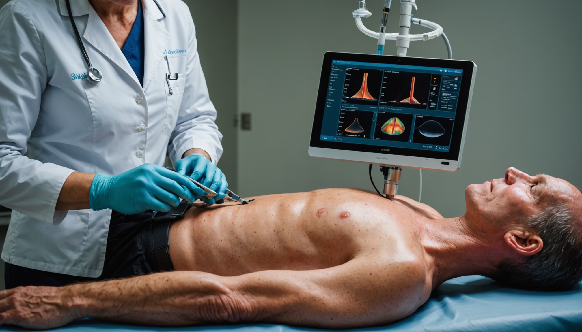
Liver cirrhosis is the end stage of chronic liver disease, in which years of inflammation and scarring replace normal liver tissue with fibrous scar tissue. The liver gradually loses its ability to function. Cirrhosis is a leading cause of liver-related death in the UK, but with the right management, disease progression can be slowed and complications reduced significantly.
What is Liver Cirrhosis?
Cirrhosis is irreversible scarring of the liver resulting from prolonged injury. As scar tissue replaces healthy liver cells, the liver's ability to perform its vital functions — detoxification, protein synthesis, clotting factor production, and bile secretion — becomes progressively impaired. There are two key stages. Compensated cirrhosis is when the liver, despite scarring, is still able to maintain most of its functions and the patient may have no symptoms. Decompensated cirrhosis is when the liver can no longer compensate and life-threatening complications develop — including ascites, variceal bleeding, hepatic encephalopathy, and jaundice.
Common Causes
The most common causes in the UK are alcohol-related liver disease (ARLD), non-alcoholic fatty liver disease (NAFLD/MASLD), and chronic hepatitis C infection. Other causes include autoimmune hepatitis, primary biliary cholangitis, primary sclerosing cholangitis, haemochromatosis, Wilson's disease, and alpha-1 antitrypsin deficiency. Careful evaluation of the underlying cause is essential — some causes are treatable (e.g. alcohol cessation, viral hepatitis treatment), which can slow or halt progression.
Symptoms and Signs
Compensated cirrhosis may be asymptomatic or cause only mild fatigue and abdominal discomfort. Signs of decompensation include jaundice (yellow skin and eyes from bilirubin accumulation), ascites (fluid in the abdomen causing distension), peripheral oedema (leg swelling), spider naevi (spider-shaped blood vessels on the skin), palmar erythema (red palms), gynaecomastia, and muscle wasting. Hepatic encephalopathy causes confusion, drowsiness, and flapping tremor (asterixis) due to ammonia accumulation. Variceal haemorrhage from oesophageal or gastric varices presents as sudden, large-volume haematemesis and is a medical emergency.
Diagnosis and Monitoring
Diagnosis involves liver function tests (LFTs), FBC, clotting studies, and imaging. Ultrasound shows a nodular, shrunken liver with altered echogenicity and may detect ascites. Transient elastography (FibroScan) measures liver stiffness non-invasively. Child-Pugh score and MELD score are used to assess severity and prognosis. All patients with cirrhosis should have six-monthly liver ultrasound and alpha-fetoprotein (AFP) for hepatocellular carcinoma (HCC) surveillance. OGD (endoscopy) is performed to screen for oesophageal varices.
Management and Complications
Management aims to treat the underlying cause, prevent decompensation, and manage complications. Alcohol cessation is the single most important intervention in ARLD. Ascites is managed with dietary sodium restriction and spironolactone (first-line diuretic) ± furosemide. Refractory ascites requires therapeutic paracentesis (with albumin infusion). Non-selective beta-blockers (propranolol or carvedilol) reduce portal hypertension and prevent first variceal bleed. Acute variceal bleeding is treated with terlipressin, antibiotics (ceftriaxone), and urgent endoscopy with band ligation. Hepatic encephalopathy is managed with lactulose (to reduce ammonia absorption) and rifaximin (to alter gut flora). All patients should be considered for liver transplantation assessment when appropriate. This article has been carefully prepared following evaluation of current NICE and BSG guidelines on liver cirrhosis.