Portal Hypertension & Varices
Understanding raised portal pressure, variceal bleeding and its emergency management
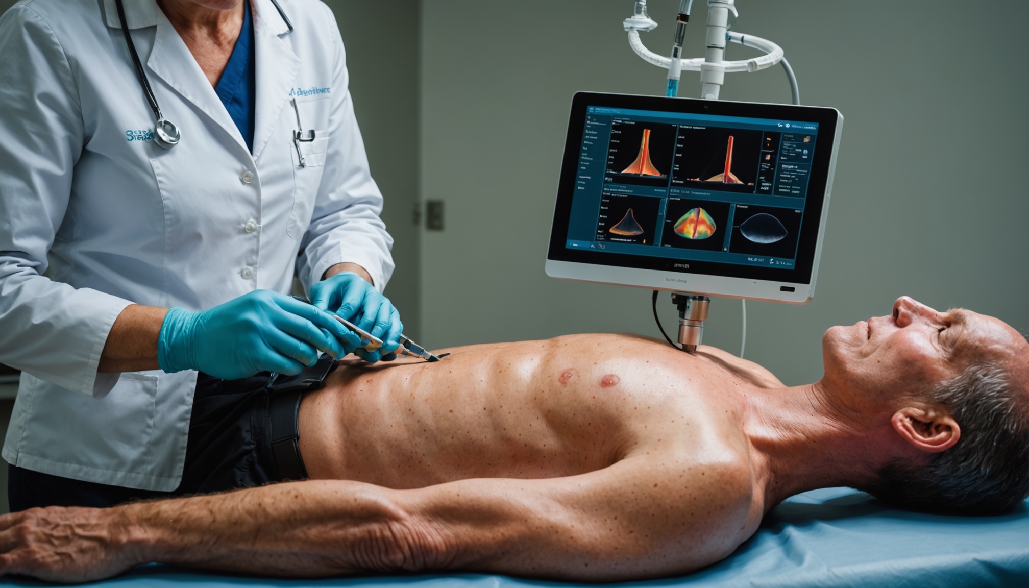
Portal hypertension is defined as an elevation of pressure in the portal venous system above 5 mmHg, with clinically significant portal hypertension occurring above 10 mmHg. It is the principal consequence of liver cirrhosis and drives its most dangerous complications — oesophageal and gastric varices, ascites, and hepatic encephalopathy. Variceal haemorrhage is a life-threatening emergency with a mortality of 15–20% per episode. Careful evaluation and systematic management of both primary prevention and acute bleeding are essential.
What is Portal Hypertension?
The portal vein collects blood from the intestines, spleen, and stomach and delivers it to the liver. In cirrhosis, fibrous scar tissue increases resistance to blood flow through the liver, raising portal pressure. When portal pressure rises above 10 mmHg, collateral vessels develop to decompress the portal system — these include oesophageal and gastric varices, caput medusae (dilated abdominal wall veins), and haemorrhoids. Portal hypertension is classified by the site of obstruction: prehepatic (portal vein thrombosis), intrahepatic (cirrhosis — the most common), and posthepatic (Budd-Chiari syndrome, right heart failure). The hepatic venous pressure gradient (HVPG) is the gold standard measure — measured during transjugular liver biopsy.
Varices — Formation and Risk of Bleeding
Oesophageal varices develop in approximately 50% of cirrhotic patients within 10 years of diagnosis. The risk of first bleeding is 25–30% at 2 years for large varices. Variceal bleeding occurs when wall tension exceeds the capacity of the variceal wall — influenced by variceal size, variceal wall tension, and portal pressure. The key risk factors for first bleed are: large variceal size (>5 mm), red wale marks on endoscopy (longitudinal red streaks on varices indicating high wall tension), Child-Pugh C liver disease, and no current primary prophylaxis. Gastric varices (particularly isolated gastric varices at the fundus — IGV1) are less common than oesophageal but bleed more severely.
Primary Prophylaxis — Preventing the First Bleed
All patients with cirrhosis should undergo upper GI endoscopy (OGD) at diagnosis to screen for varices. If no varices: repeat OGD every 2–3 years (or annually if decompensated). Small varices without high-risk features: repeat OGD in 1–2 years. Large varices or small varices with high-risk features: start primary prophylaxis. Non-selective beta-blockers (NSBBs) — propranolol or carvedilol — are the pharmacological treatment of choice. They reduce portal pressure by decreasing cardiac output and splanchnic vasoconstriction. Carvedilol 6.25 mg OD is increasingly preferred over propranolol due to superior portal pressure reduction. Endoscopic variceal band ligation (EVBL) is equally effective and preferred if NSBBs are contraindicated or poorly tolerated. Nitrates are not recommended for primary prophylaxis.
Acute Variceal Haemorrhage — Emergency Management
Acute variceal bleeding is a medical emergency. Mortality is 15–20% per episode. The management follows a structured approach. Resuscitation first: two large-bore IV cannulae, crossmatch, IV fluid resuscitation (avoid over-transfusion — target Hb 70–80 g/L using packed red cells as over-transfusion raises portal pressure and worsens bleeding). Vasoactive drugs: terlipressin 2 mg IV every 4 hours (reduces portal pressure by splanchnic vasoconstriction) — start immediately and continue for 5 days. Antibiotic prophylaxis: IV ceftriaxone 1 g daily for 5–7 days — reduces bacterial translocation, SBP risk, and mortality. Urgent OGD within 12 hours (ideally within 6 hours): endoscopic band ligation of oesophageal varices is the treatment of choice. Balloon tamponade (Sengstaken-Blakemore tube) as a bridge to definitive treatment if endoscopy fails or unavailable. This article has been prepared following evaluation of NICE, BSG, and EASL guidelines on variceal haemorrhage.
Secondary Prophylaxis and TIPS
After surviving a variceal bleed, the risk of rebleeding is 60% within 1–2 years without treatment. Secondary prophylaxis combines endoscopic band ligation (every 2–4 weeks until obliteration) plus non-selective beta-blockers. TIPS (Transjugular Intrahepatic Portosystemic Shunt) is used for patients who fail secondary prophylaxis or have recurrent bleeding — it creates a channel between the portal and hepatic veins, dramatically reducing portal pressure. Pre-emptive TIPS within 72 hours of index variceal bleed in high-risk patients (Child-Pugh B with active bleeding at OGD, or Child-Pugh C) significantly reduces rebleeding and mortality. Patients successfully treated for varices should continue HCC surveillance and optimisation of underlying liver disease.